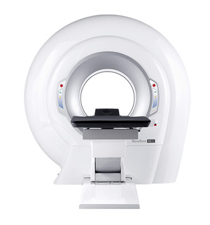
NEWTOM
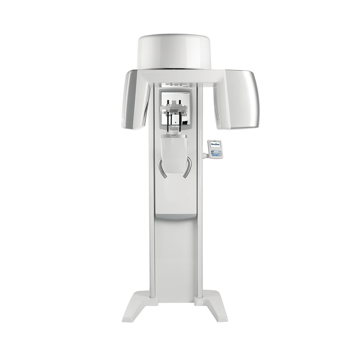
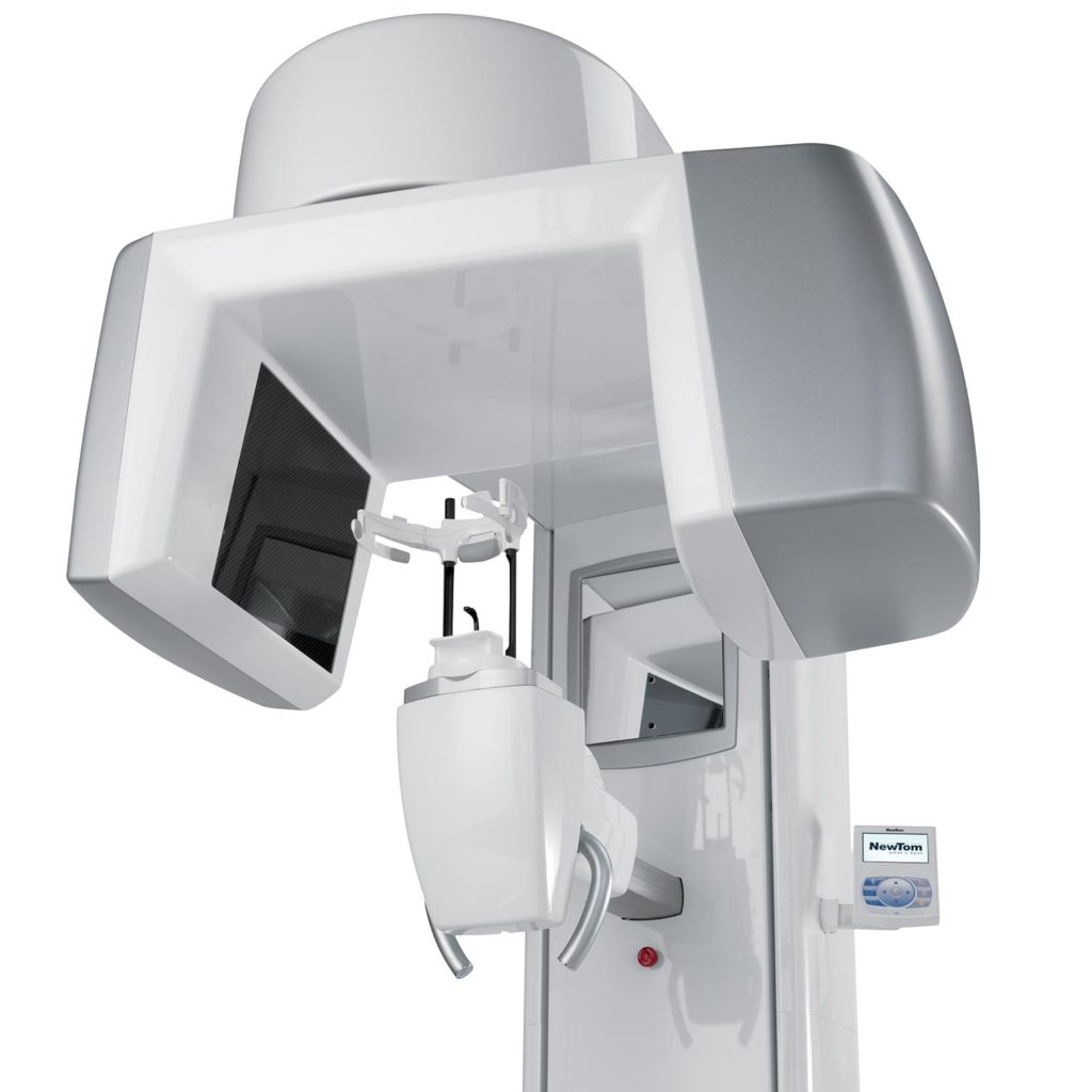
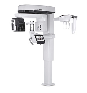
VGi evo
EXPANDED.VISION
From Newtom’s research and innovation, the most complete maxillofacial/ent CBCT.
The technologically advanced elements that form the innovative image chain of VGi evo carry the performance of CBCT devices to a new extraordinary level.
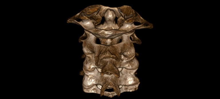
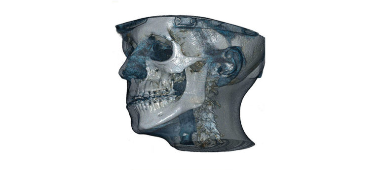
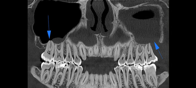
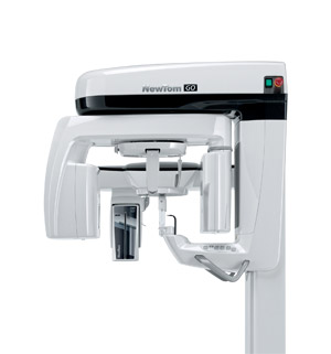
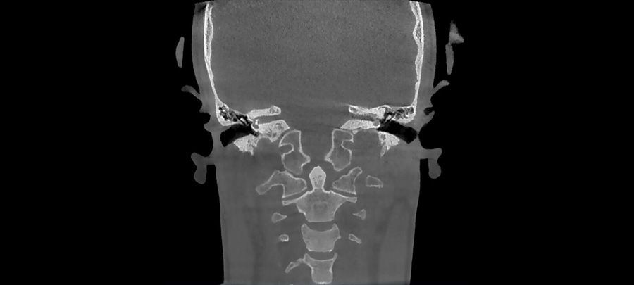
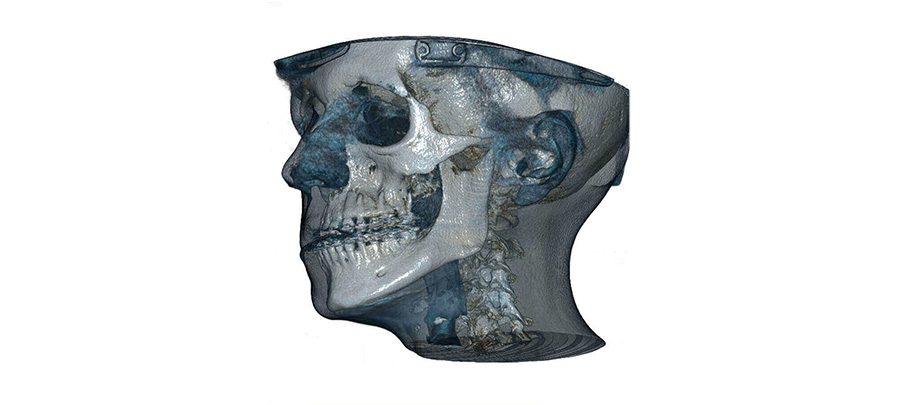
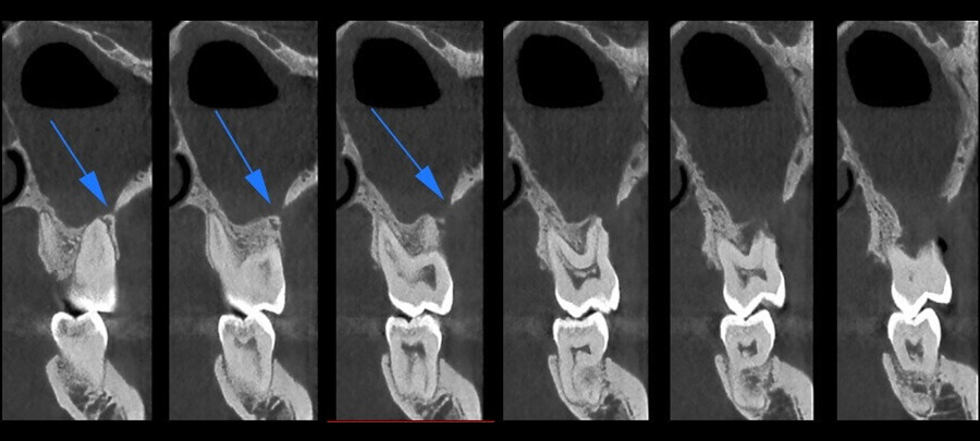
EXTENDED 3D DIAGNOSTICS
Complete FOV range for perfect 3D volumes in all situations.
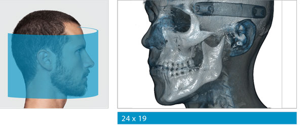
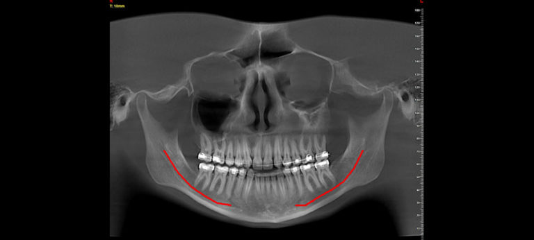
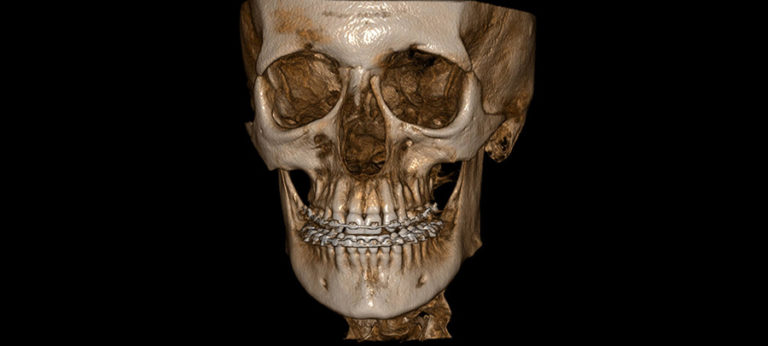
WIDE-RANGING FOVs
The most wide-ranging FOVs allow, with a single scan, to see complete images of the maxillofacial area for orthodontic applications, orthognathic and maxillofacial surgery.
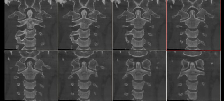
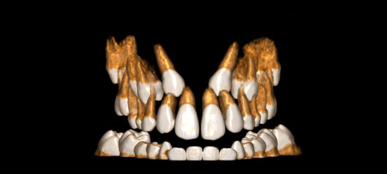
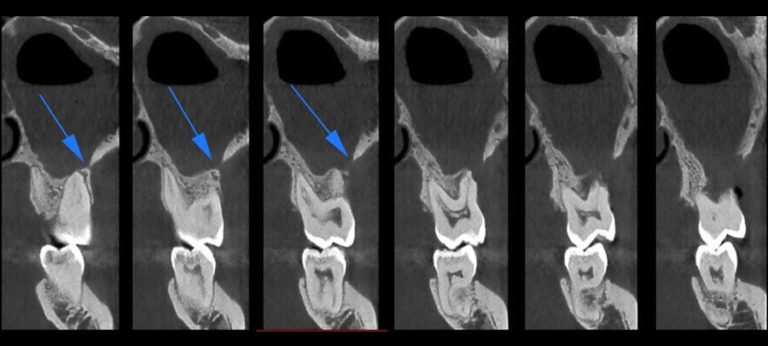
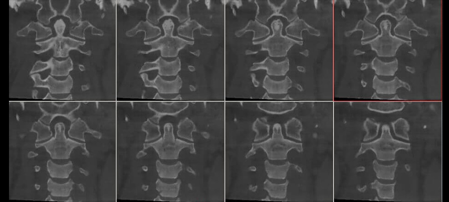
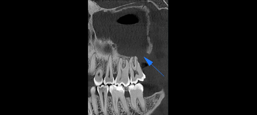
MEDIUM-SIZE FOVs
Medium-size FOVs are indicated for ENT (otorhinolaryngology) and TMJ (Temporomandibular Joint) applications or full dentition examination and implant planning.
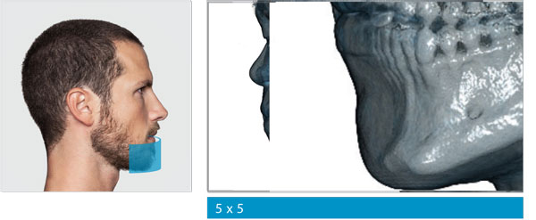
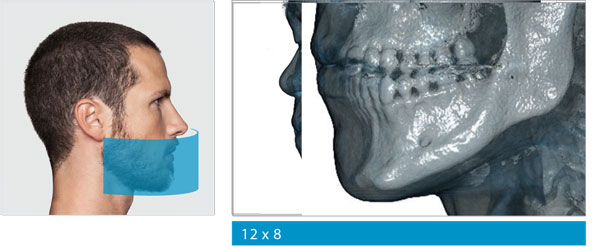
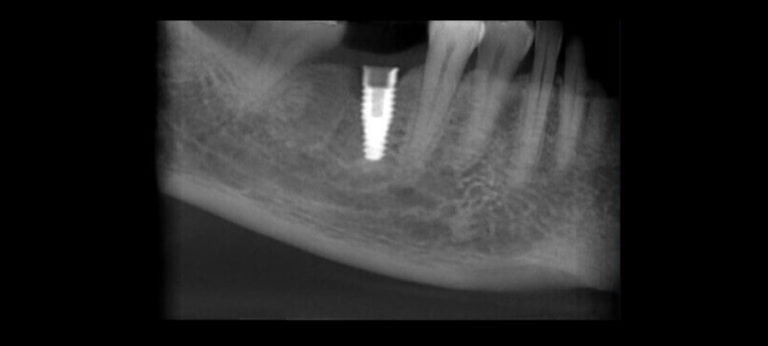
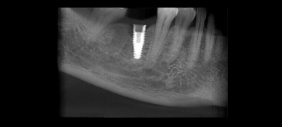
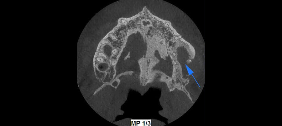
SMALL FOVs
Use of small FOVs is indicated for ENT, endodontic, periodontic and implantology examinations on specific user-selected regions.
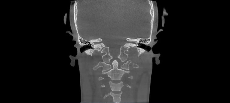
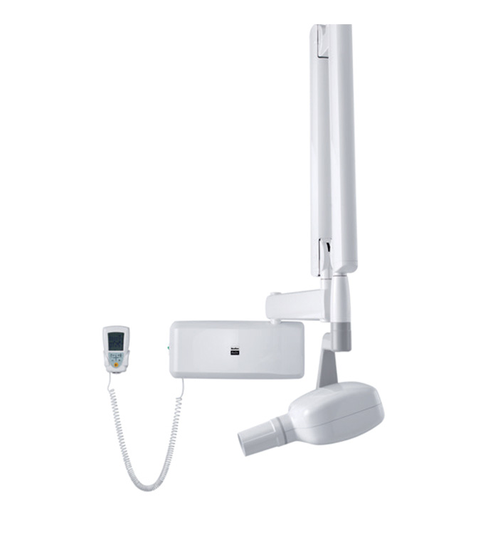
VERSATILE 2D IMAGING
Panoramic and cephalometric examinations for a precise and complete view.
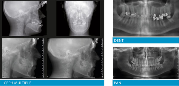
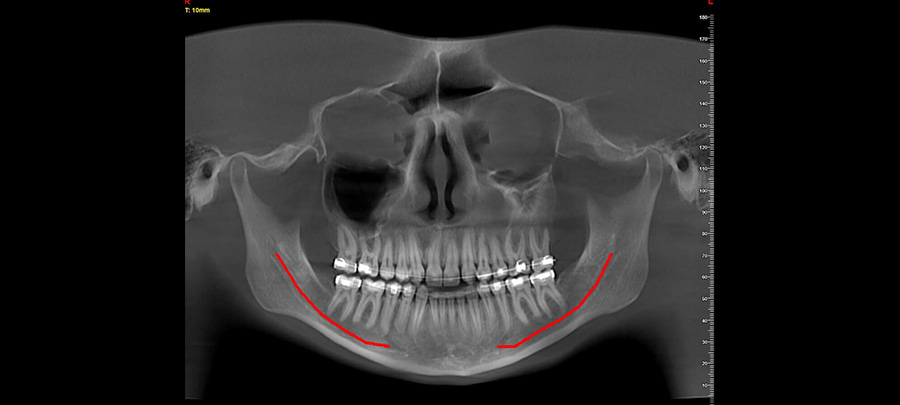
Sharp 2D – TELERADIOGRAPHIC AND PANORAMIC SCANS
Exclusive function to create a dataset of images from Panoramic and Teleradiographic (AP, PA and LL) scans in a single examination.
Compared to the panoramic scan-like coronal reconstructions (panorex) conventionally obtained with CBCT, images produced with Sharp 2D maintain the same magnification and orthogonality ratios; hence, the same clinical evaluation principles of conventional panoramic views apply.
Latero-Lateral and Antero-Posterior teleradiographic scans can be used to perform cephalometric examinations and orthodontic rehabilitation.
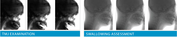
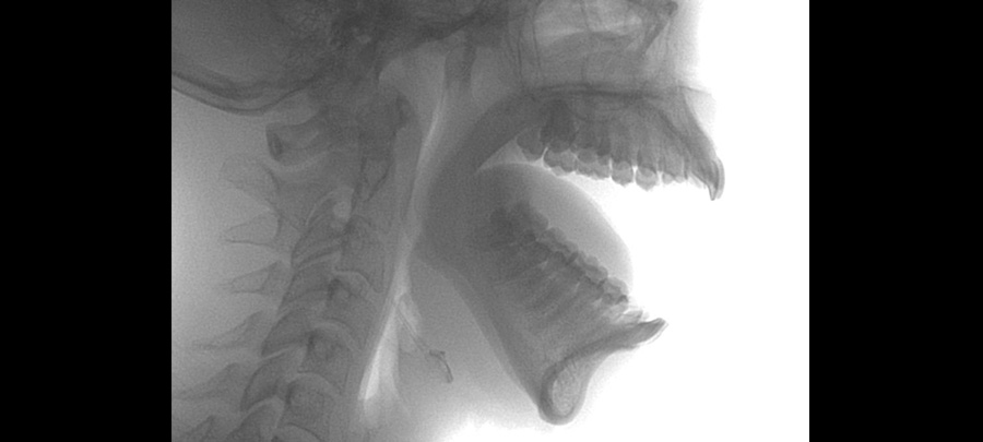
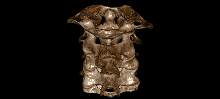
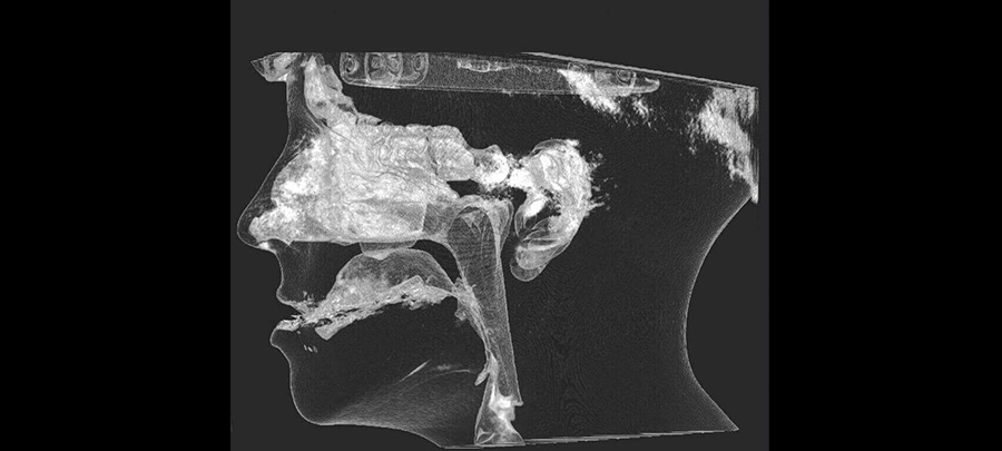
CineX – DYNAMIC IMAGES
The innovative CineX function, which is provided with a 17 x 19 cm filming area, allows to investigate moving internal anatomical structures (e.g., swallowing, saliva ducts, TMJ disc, cervical vertebrae) by acquiring a sequence of X-rays in video format with AP, PA or LL projections.
The videos obtained can then be directly consulted with the NNT software, with the NNT Viewer or exported and viewed with third party applications.

EXCEPTIONAL OPERATOR AND PATIENT COMFORT
Effective communication
Precise diagnosis and complete planning of treatment ensure effective communications between specialist physician and patient, an essential requisite to learn about the treatment in a safe and trusted setting.
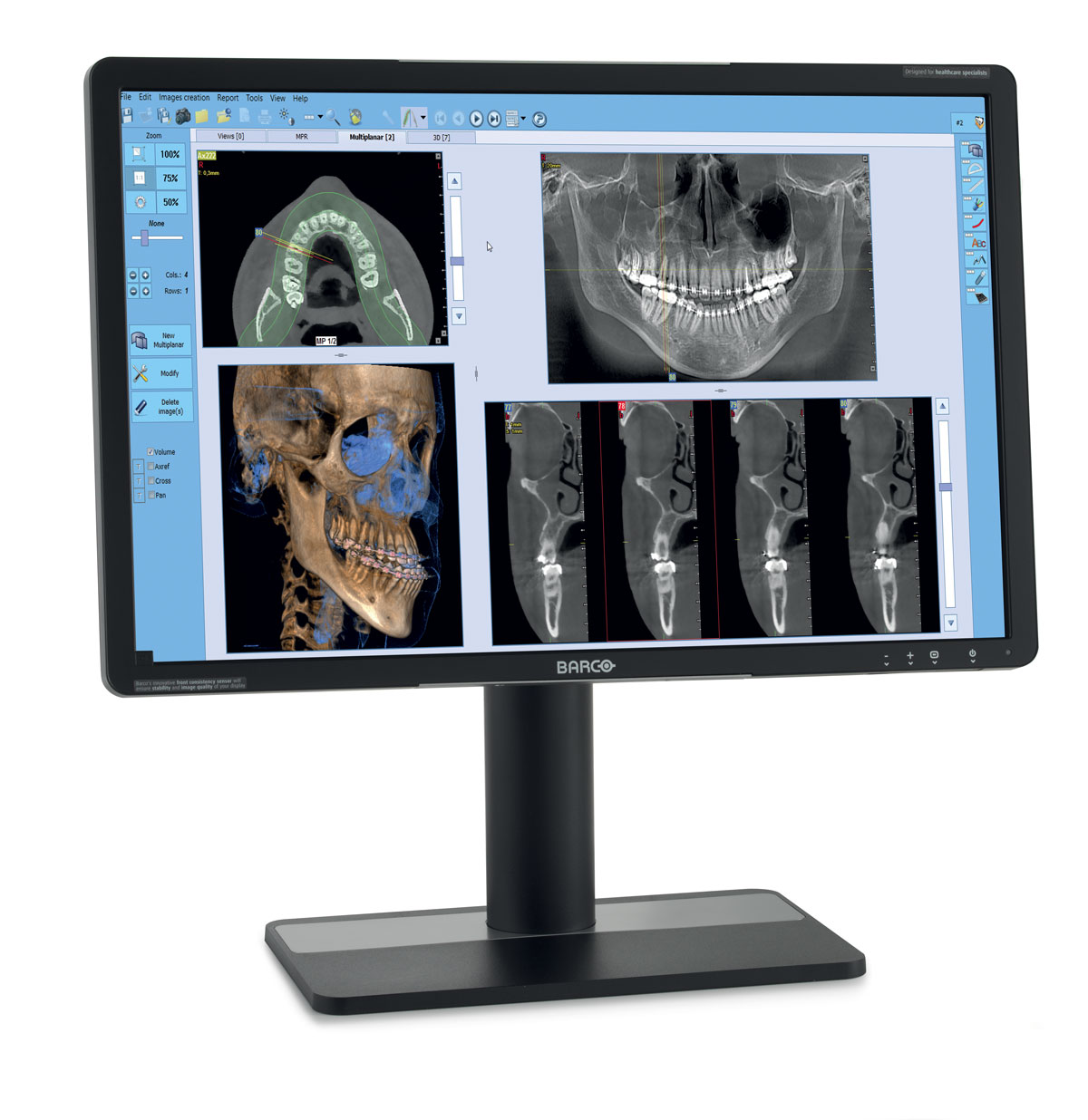
NNT
TECHNOLOGICAL HEART
Technologically advanced software for 2D and 3D imaging. With just a few simple steps our NNT software can process data acquired during the scan to create a vast array of images, which provide detailed information about patient anatomy.
They can subsequently be saved in a report or distributed with the Viewer version of the software. NNT also provides different application modes specifically intended for implantology, endodontics, periodontics, maxillofacial surgery and radiology.


Cefla Medical Equipment is considered Europe’s number one dental unit manufacturer.