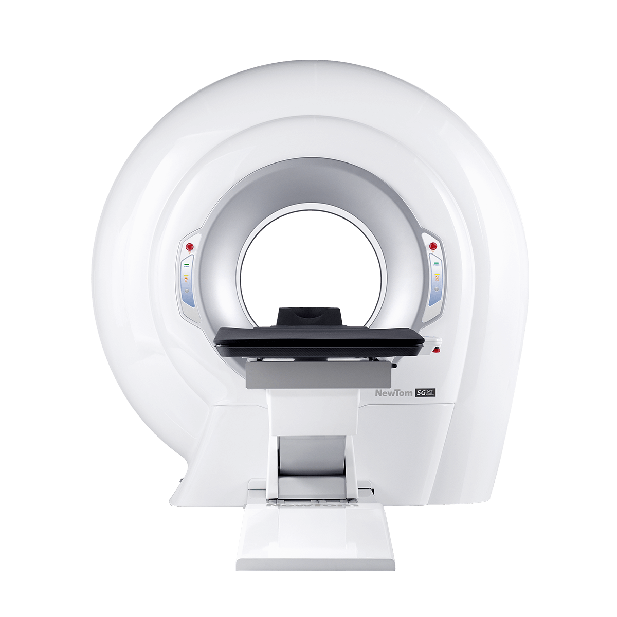
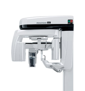
NEWTOM 5G XL
EXTRA.VISION
The ultimate 3D
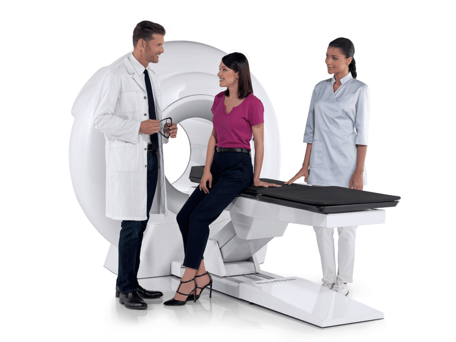
The only CBCT with lying down patient positioning. excellent image quality with a device that features outstanding diagnostic potential.
5G XL is the innovative device, featuring lying down patient positioning, able to offer high resolution volumetric images with extra low X-ray doses. Top quality CBCT for new medical applications.
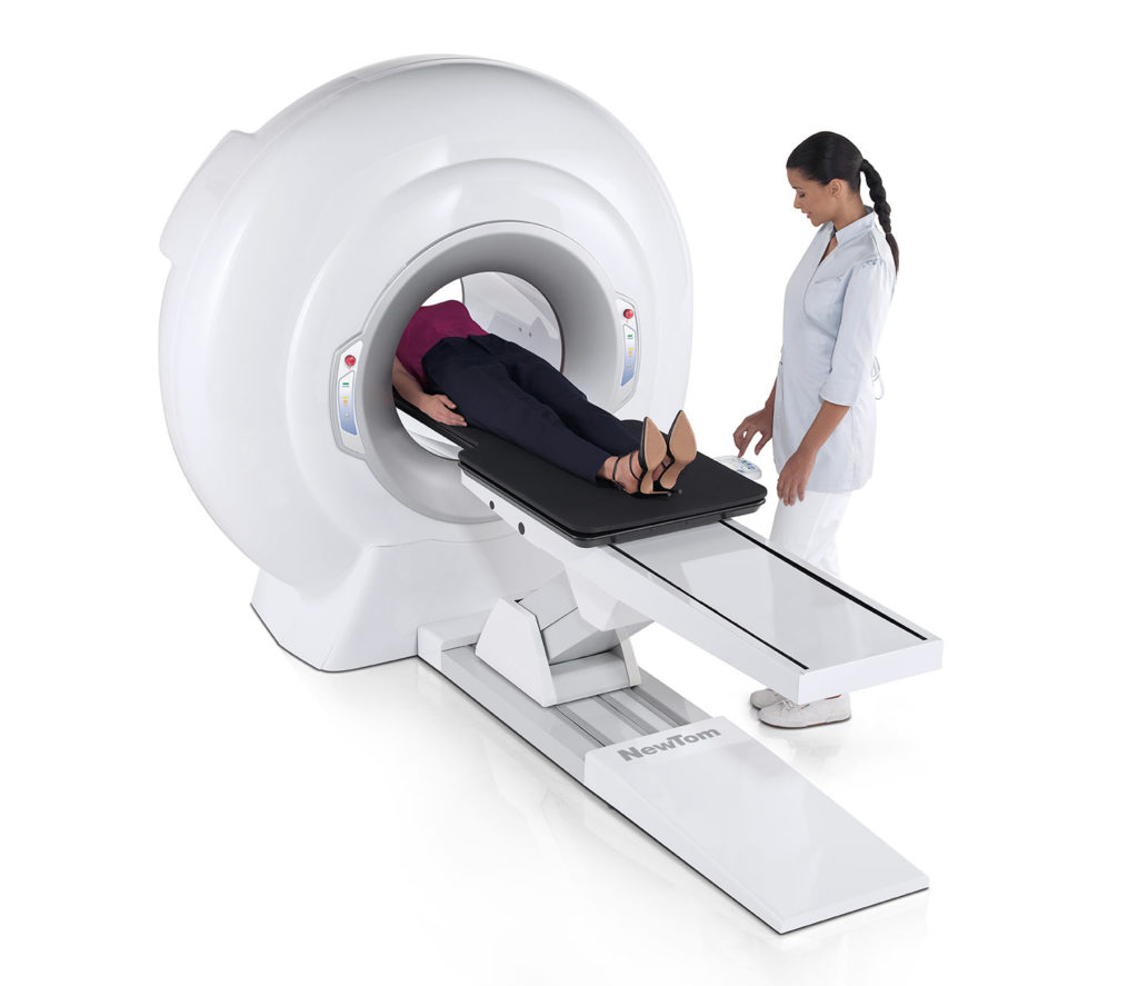
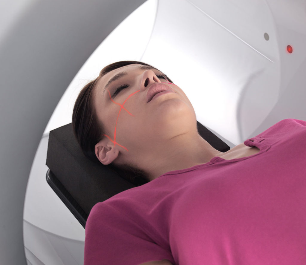
OPTIMAL LYING DOWN POSITIONING
User-friendliness, maximum stabilization and quality for diagnoses using new medical applications.
5G XL is the only CBCT device available on the market with patient in a lying down position. The motor-driven patient table made of carbon fiber, which can be controlled from the on-board console or from the PC, allows to adapt the examination to any image acquisition need with patient lying down in a prone or supine, cranial-caudal or caudal-cranial position. The positioning and lock device has been specifically designed for the various dental and medical disciplines.
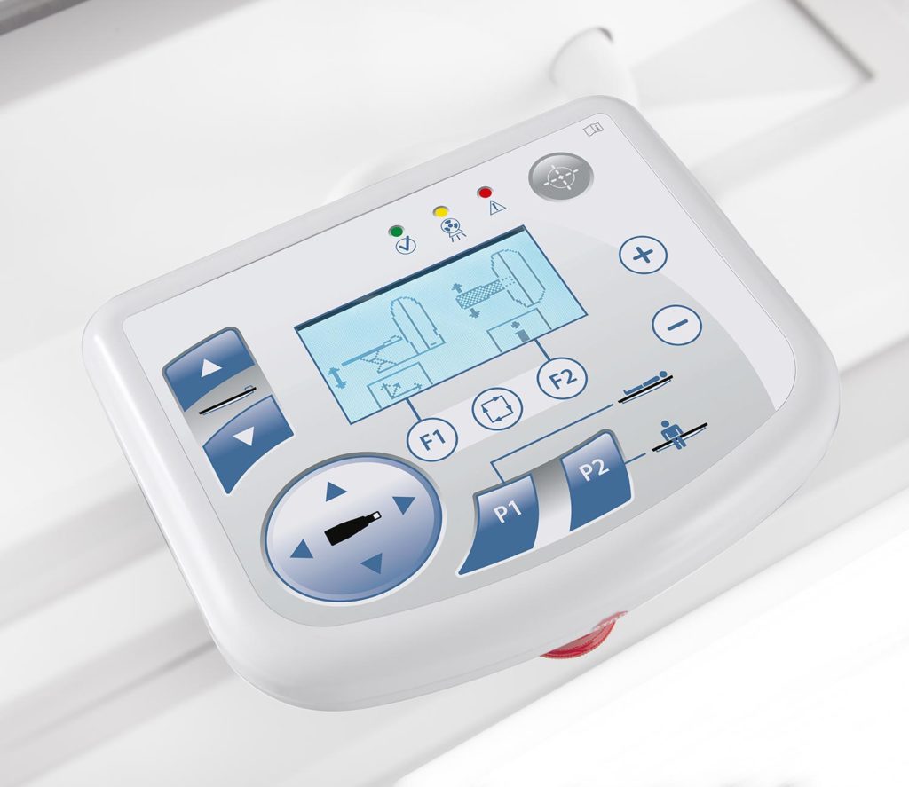
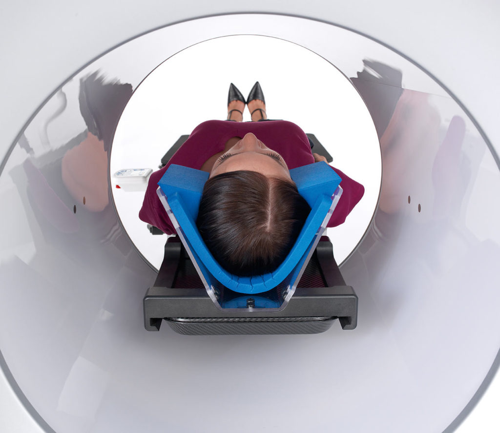
On board console
The on-board console offers a user-friendly interface to easily move the patient table along three axes, and to activate the alignment lasers by defining the exact reference points of the area of interest.
Assisted alignement
The operator directly performs assisted alignment from the workstation via two scout images for automatic adjustment of the motor-driven patient table.
LOW X-RAY DOSE. WELLBEING AND SAFETY ARE CENTRAL TO NEWTOM RESEARCH.

ECO Scan
Low emission up to 0.9 seconds of emission for standard examinations. The ECO Scan protocol is ideal for post-surgery follow-ups and paediatric applications.
SafeBeam™
The exclusive SafeBeamTM technology eliminates the risk of exposing the patient to an unnecessarily high dose by automatically adapting radiation levels to suit the patient’s anatomical characteristics.
Ray2D
The Ray2D function allows to perform a preliminary low dose 2D X-ray examination, which can be followed, if necessary, by a high resolution 3D examination only of the area of interest, for in-depth diagnostics.
SUPERIOR STANDARD 3D EXAMINATIONS WITH A DEVICE DESIGNED FOR EXCELLENT PERFORMANCE.
TECHNOLOGY
The powerful generator
The powerful generator
The powerful generator with rotating anode and smaller focal spot optimises performance, adapting the emission to the specific needs of the examination.
Flat panel sensor
A large flat panel sensor with a high signal-noise ratio improves image quality, expanding 3D and 2D diagnostic capacity.
PERFORMANCE

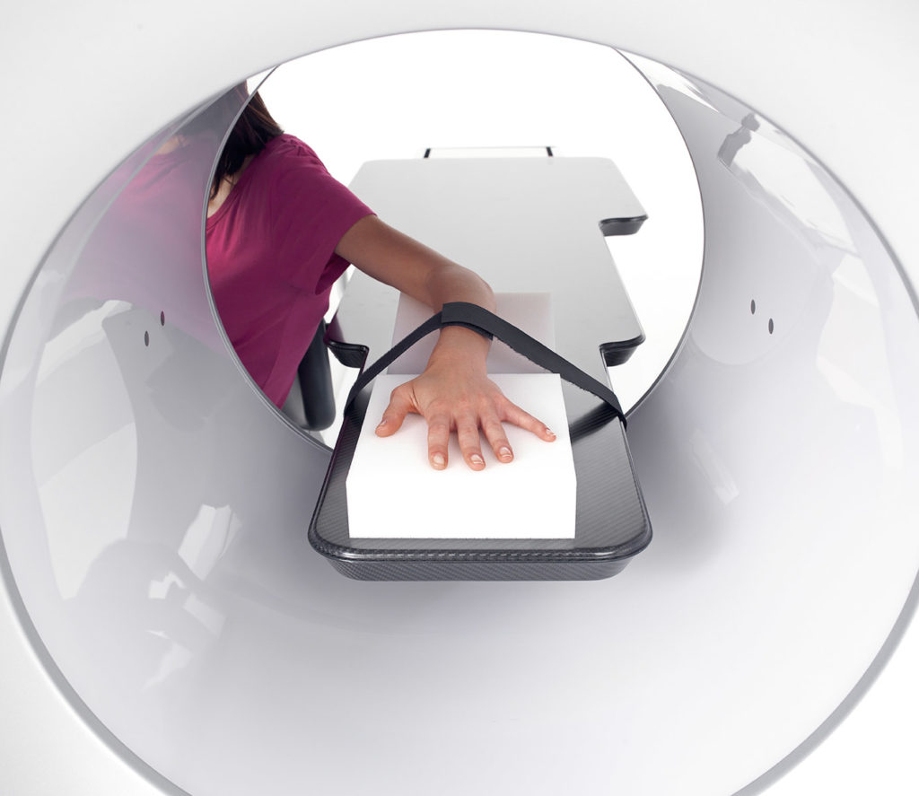
EXtra FOV Vision
The innovative eXtra FOV function allows to examine longitudinal anatomical parts. FOV 3D can be set from a minimum of Ø6 x h6 cm, up to a maximum native diameter of 21 cm or a height of 22 cm.
360° reconstruction
The 360° scan acquires the entire volume with a single rotation. 5G XL rapidly generates a complete dataset of axial, coronal and sagittal images as well as 3D renderings.

HiRes analysis
Clear and detailed very high resolution images to see bone micro-fractures or examine anatomical districts with micrometric details.
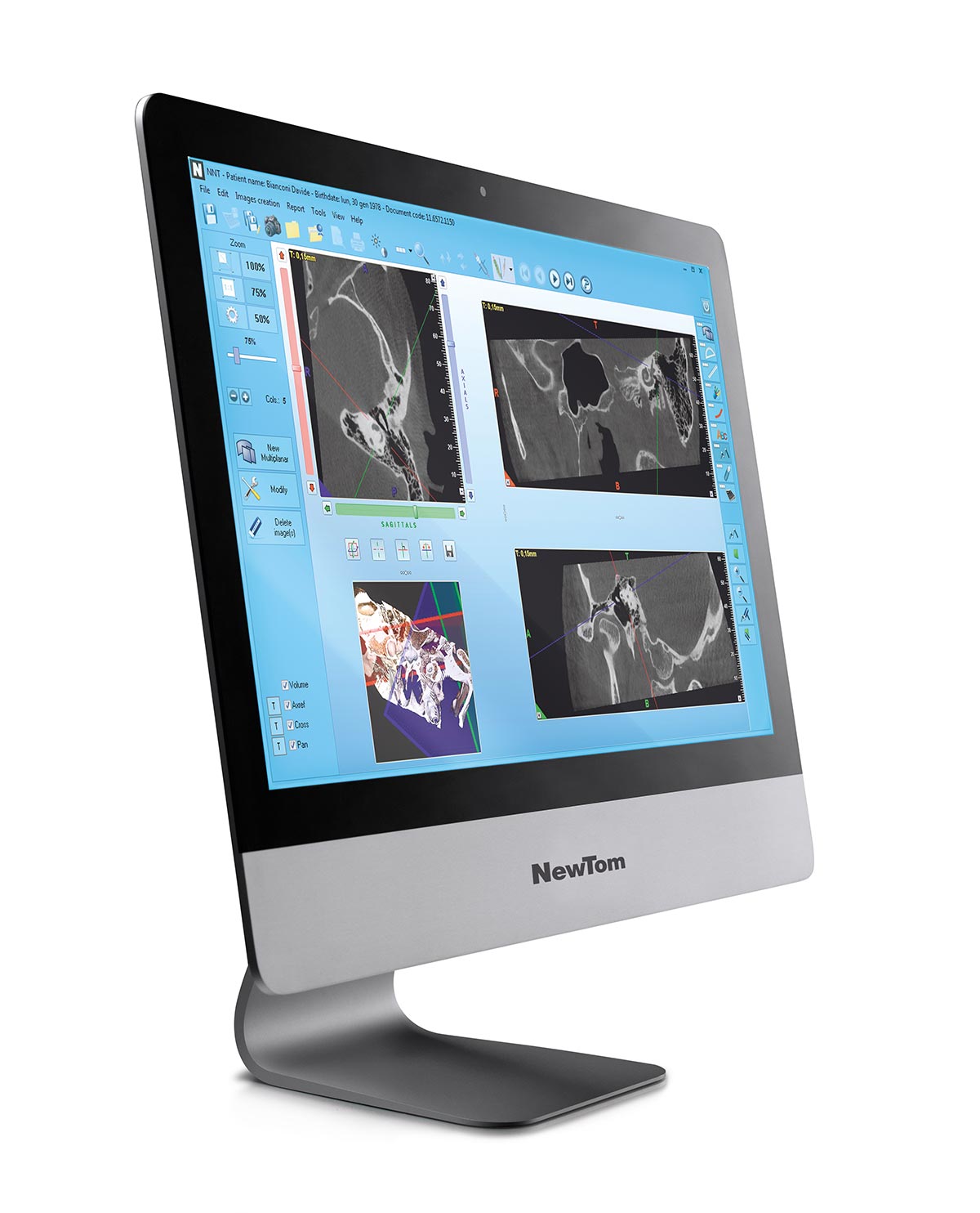
NNT, THE SOFTWARE FOR EVERY SPECIALISED NEED.
The versatile and powerful imaging software to perform the examination, process data and share the diagnosis.
SPECIAL TOOLS
Ray2D
With the innovative Ray2D function, 5G XL generates 2D X-ray images (18 x 19 cm) that are perfect for both preliminary and post-surgery follow up examinations. It is possible to repeat the investigation from various angles to select the image with the best perspective.
CineX
5G XL offers the exclusive CineX function to investigate moving anatomical structures, such as saliva ducts and joint mobility. This advanced technology uses a sequence of X-ray images to create an 18 x 19 cm format video that can also be exported to standard video format.
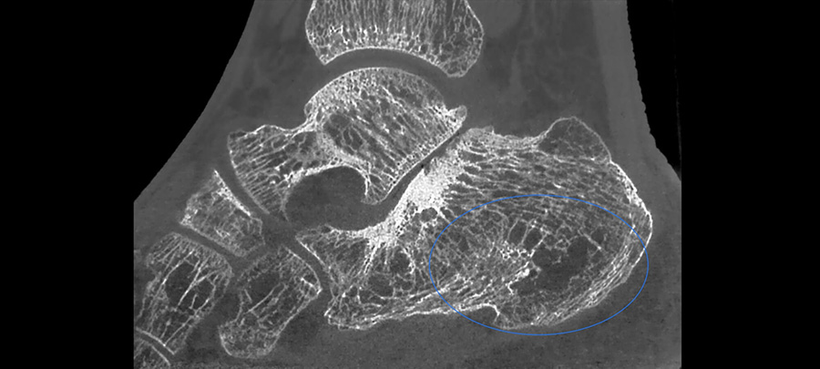
IMPLANT SITE ASSESSMENT
Estimates bone density in a potential implant site, with Misch scale classification, to correctly plan treatment.
MEASURING AIRWAY VOLUME
Quantitative measurement of upper airway space is essential to diagnose respiratory diseases and sleep apnea.
2D AND 3D MEASUREMENTS
The possibility of measuring distances on 2D sections or with 3D rendering to verify any joint problems.
CLINICAL CASES
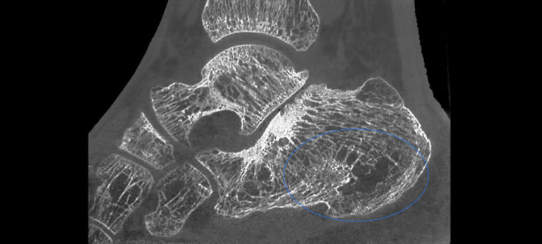
ANKLE
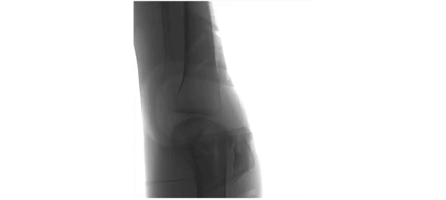
ELBOW
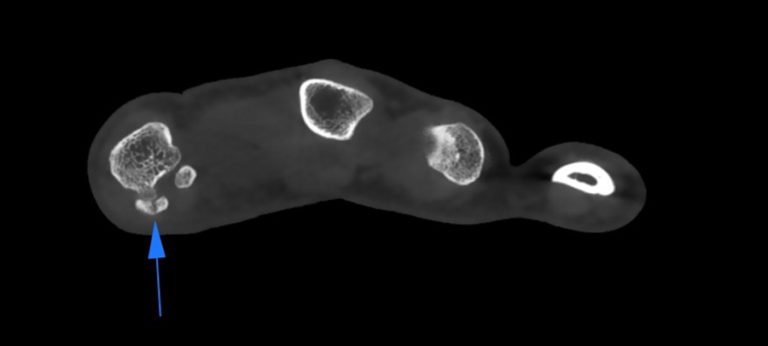
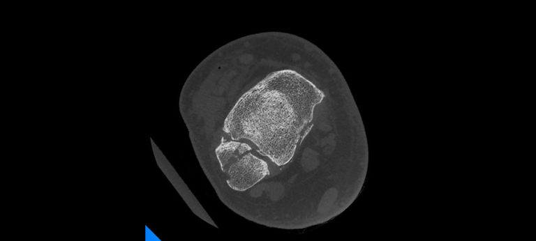
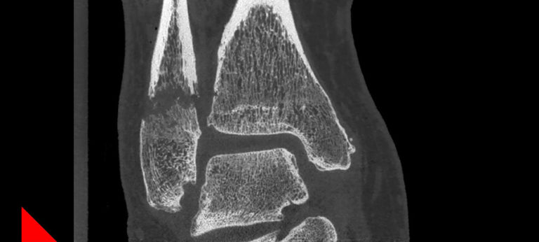
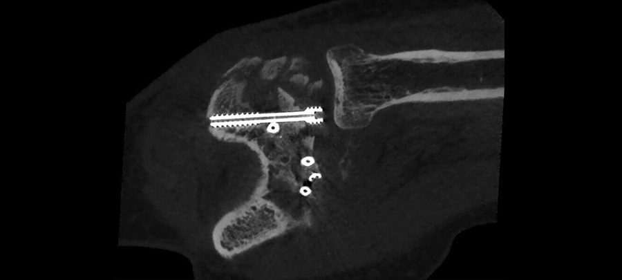
FOOT
HAND
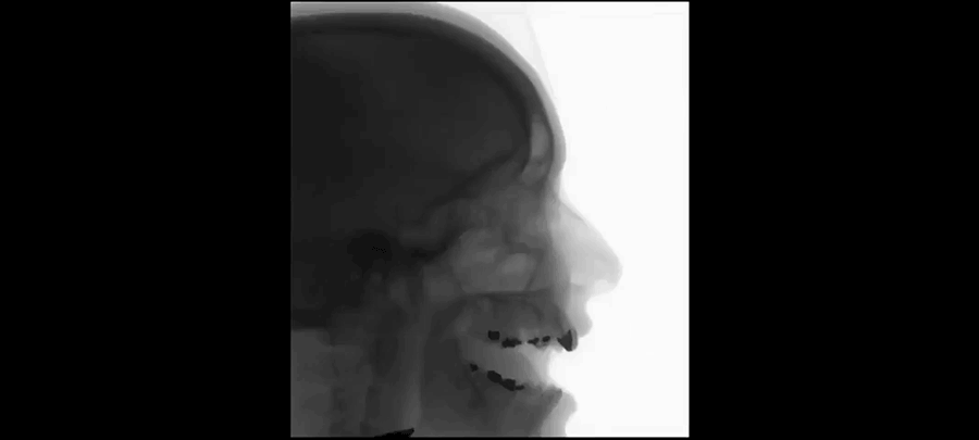
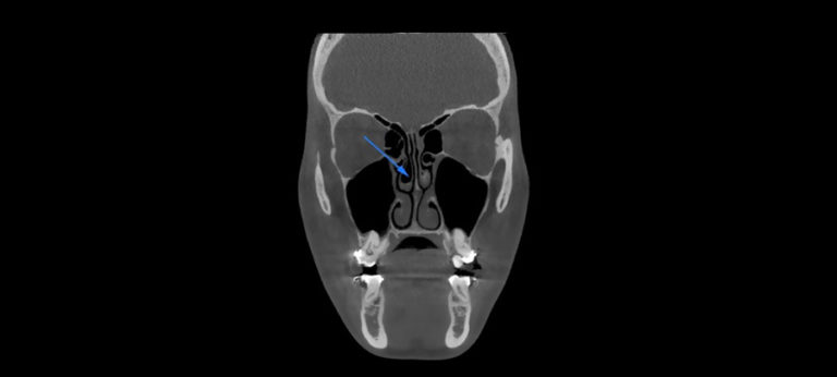
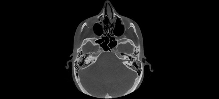
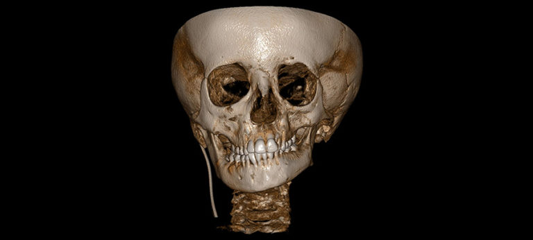
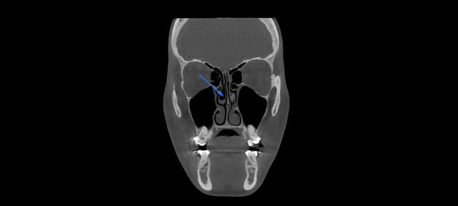
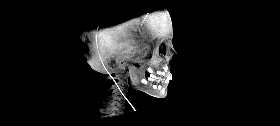
HEAD
Ceph
Mxf
Ent
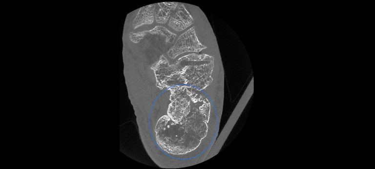
KNEE
TIBIA-FIBULA
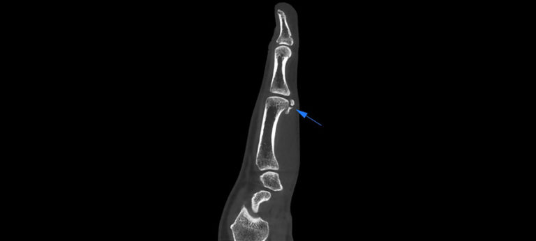
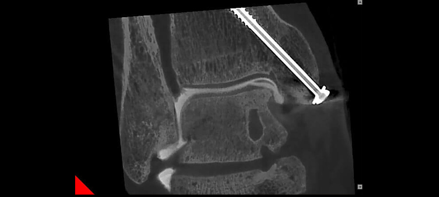
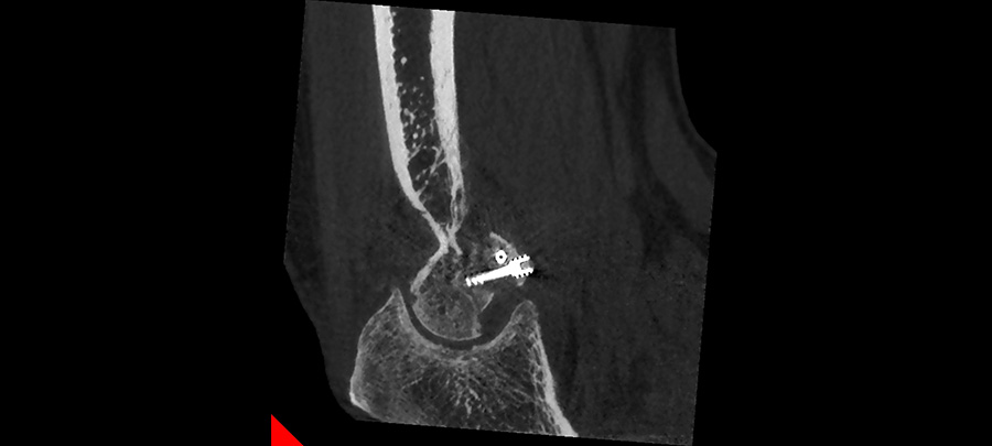
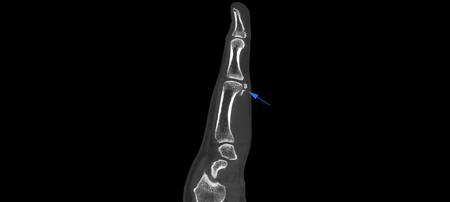
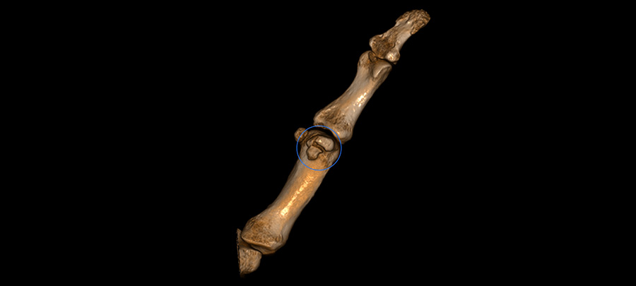
WRIST


HEAD
DEGLUTITION – Dinamic CineX
Ceph
DEGLUTITION – Dinamic CineX

KNEE
KNEE JOINT
Dinamic CineX
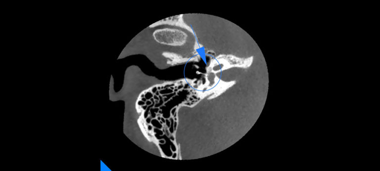
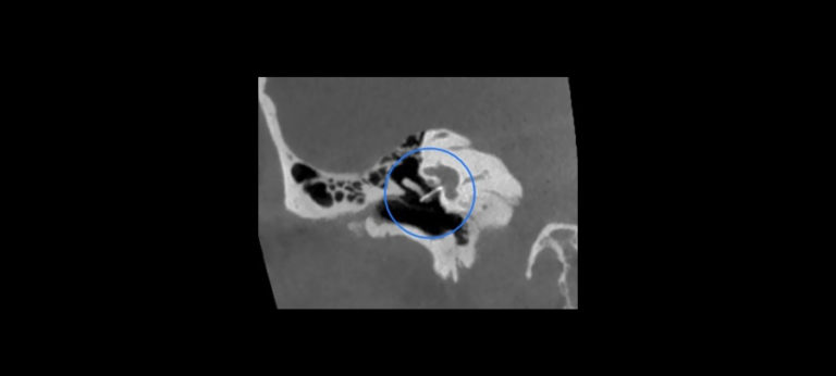
WRIST
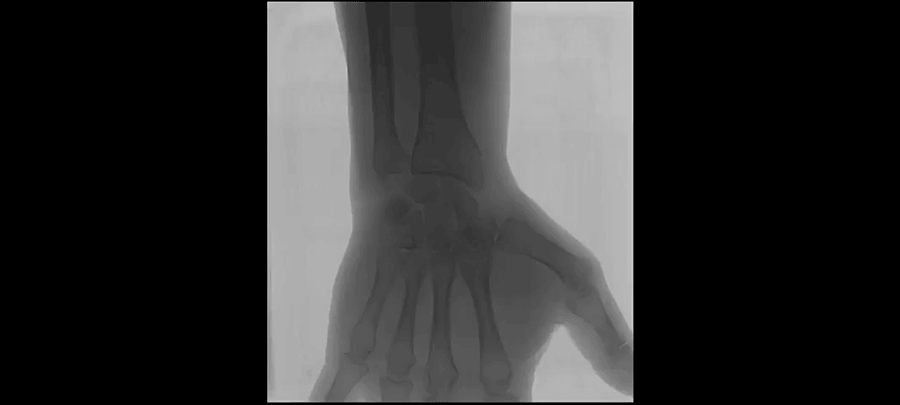
RADIUS STYLOID PROCESS FRACTURE
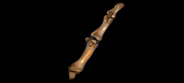
3D MSK
Cefla Medical Equipment is considered Europe’s number one dental unit manufacturer.