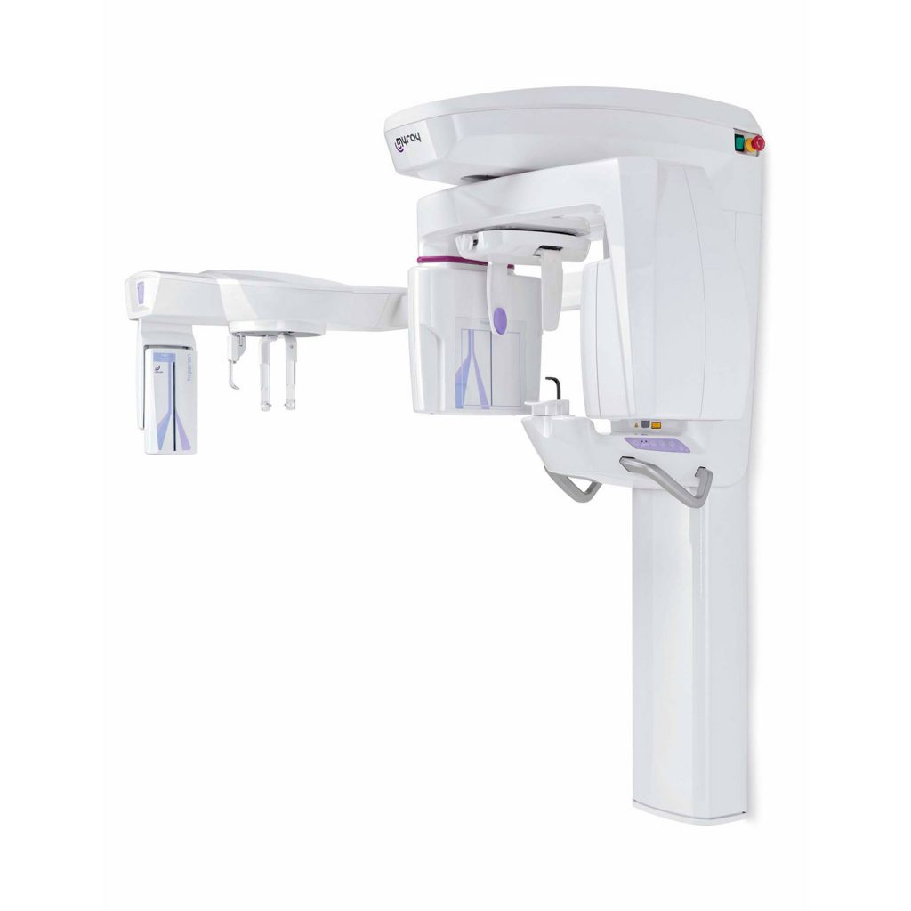
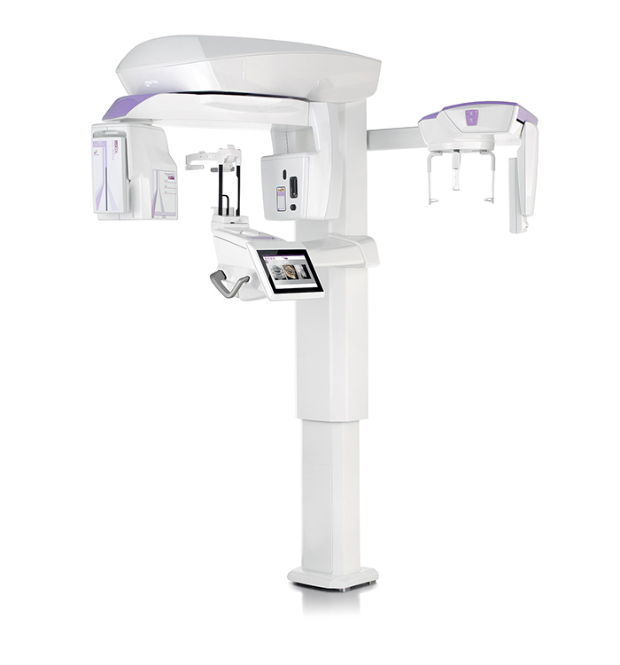
MYRAY HYPERION X5
3D/2D CEPH Imaging
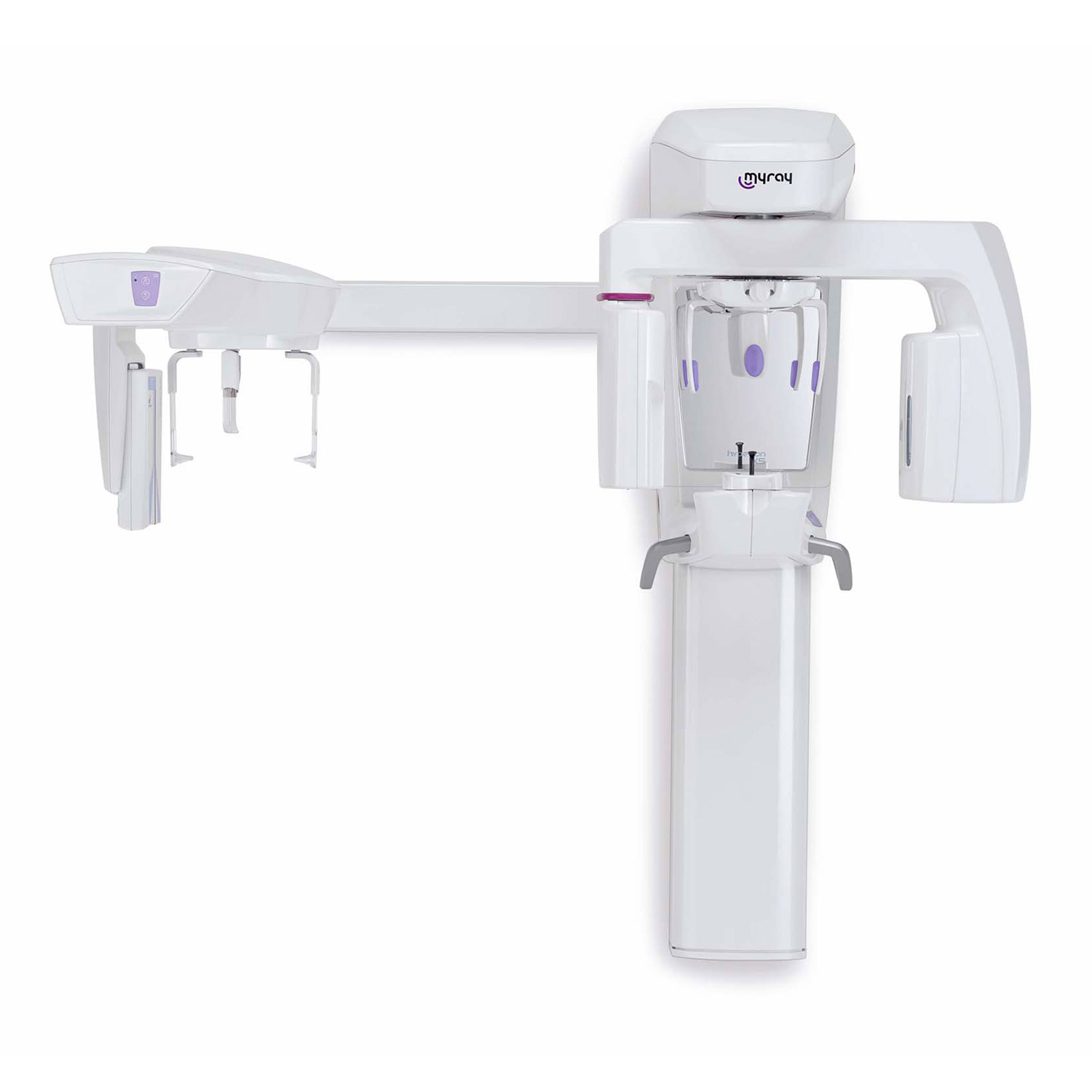
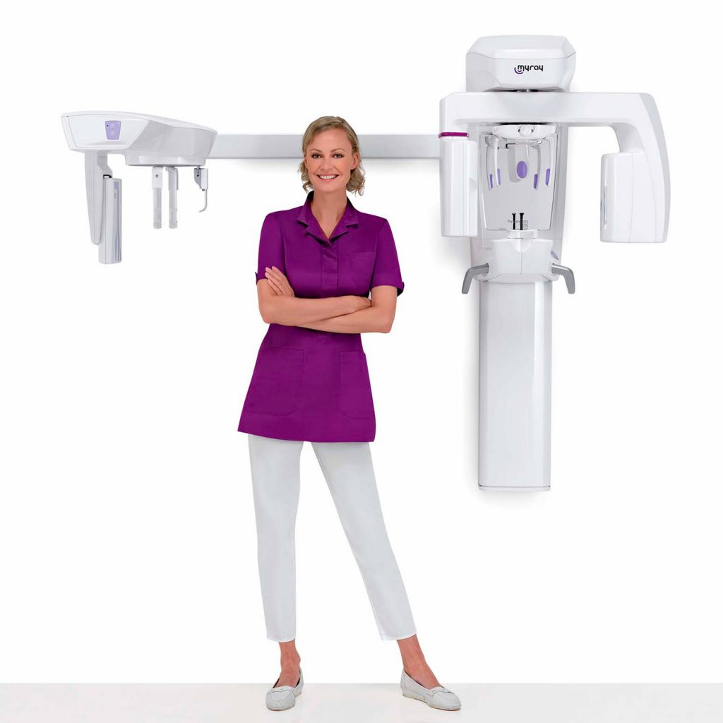
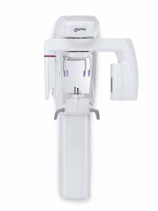
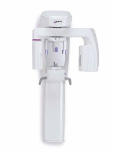
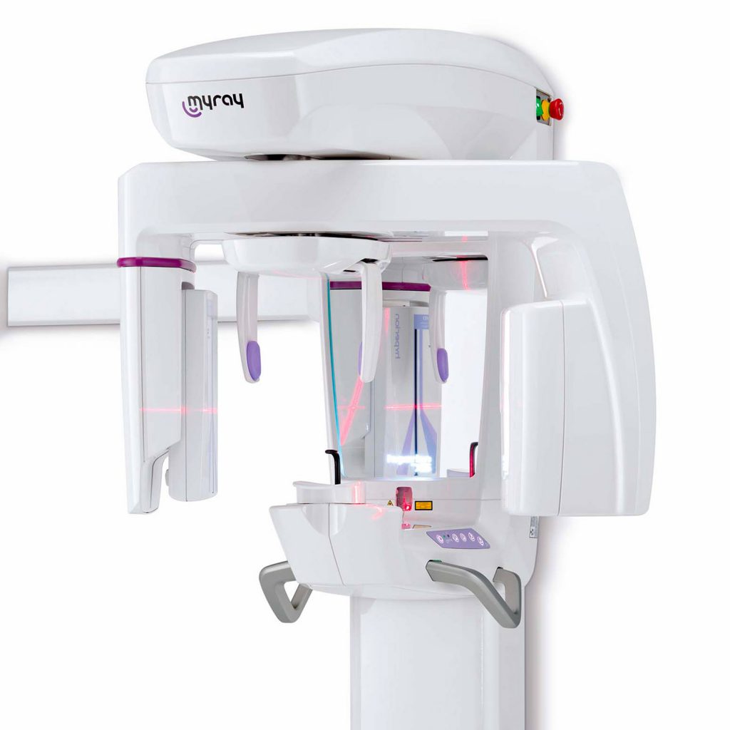
The smallest 3D/2D suspended system in the world evolves to integrate teleradiographic examinations as an extra option. Innovative design, flexibility and user-friendliness. Out of our experience comes the best solution for every dentist.
CONTINUOUS INNOVATION
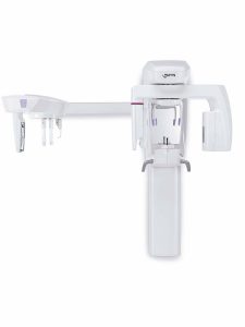
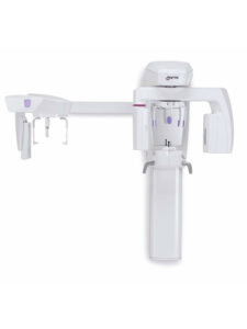
Hyperion X5 evolves to let the dentist choose a Ceph application, which can also be retrofitted.
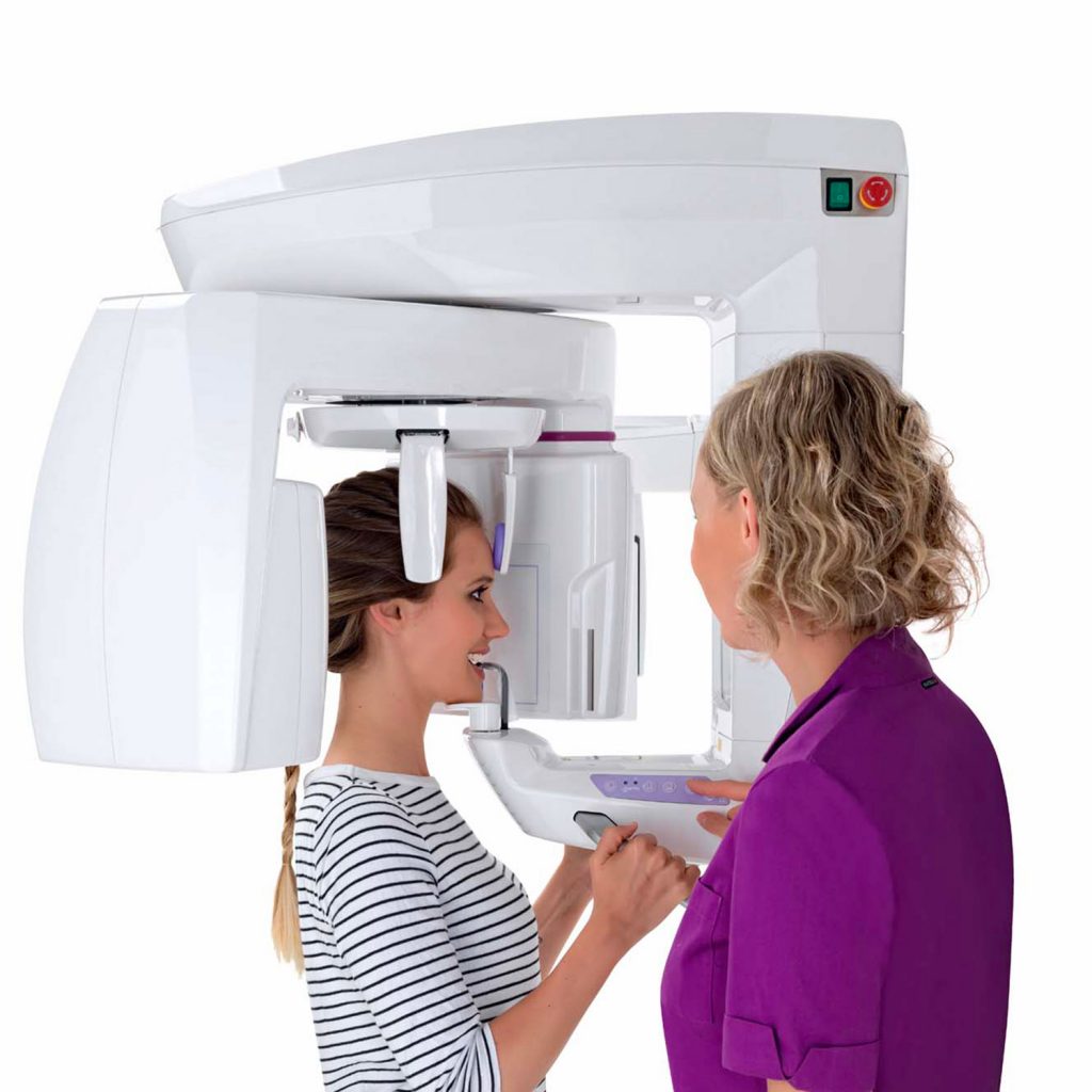
Quick and easy to use throughout the examination, this system ensures high resolution 3D and 2D images and low emission times plus fast data processing for real time diagnosis and improved patient communication.
The new virtual console streamlines capturing procedures and introduces new protocols for volumetric examination of maxillary sinuses and orthogonal panoramic images. Thanks to the automatic servo-controlled movements of the 3D sensor block, short examination times ensure a consistently positive experience.
DESIGNED TO SATISFY YOUR EVERY NEED
A complete family of dental imaging solutions for all dental surgeries.
Designed for surgeries that require three-dimensional diagnostic potential, the 3D/2D-configuration Hyperion X5 offers just the right solution and simultaneously provides excellent 2D performance.
The optional integration of the teleradiographic arm further boosts the diagnostic capacity.
DIAGNOSTICS FLEXIBILITY
Flexible, efficient, fast. Hyperion X5 – designed to deliver the best results in minimum time with limited doses. It displays 2D and 3D images packed full of details to produce effective and safe diagnoses.
- MultiPAN system
- Extremely high definition 3D (80 μm)
- Clever collimation
- Real-time diagnostics
- Secure & Safe
FULL CEPH
The updated Hyperion X5 Ceph teleradiographic system features programs for every diagnostic need. Ultra-high quality images, extremely short scan times and low irradiation doses: the very best cephalometric technology, all in the most compact unit the market has to offer.

MAXI FLEX
From 2D to 3D, all the diagnostic potential you need.
From adults to children, in just a few simple steps. Adapts field of view and doses to actual diagnostic requirements. Intelligent MultiFOV collimation, from the entire dentition (10×10 cm) to just a small portion (6×6 cm). Users can select, according to diagnostic requirements, between HD (80 μm) or low-dose QuickScan (160 μm) protocols.

MULTI VISION
Advanced 2D image processing system, equipped with a MultiPAN feature able to generate in a single scan, with the same exposure levels as in traditional panoramic imaging, 5 different focussing layers from which to select the most appropriate one for your diagnostic needs. Highly useful for analysing patients with complex anatomies and/or correcting post-capture patient positioning virtually.
QUICK SCAN
Available for both 2D and 3D scans, QuickScan protocols minimise scan times and protect patient health by reducing X-ray doses.
ALL THE POTENTIAL OF 3D
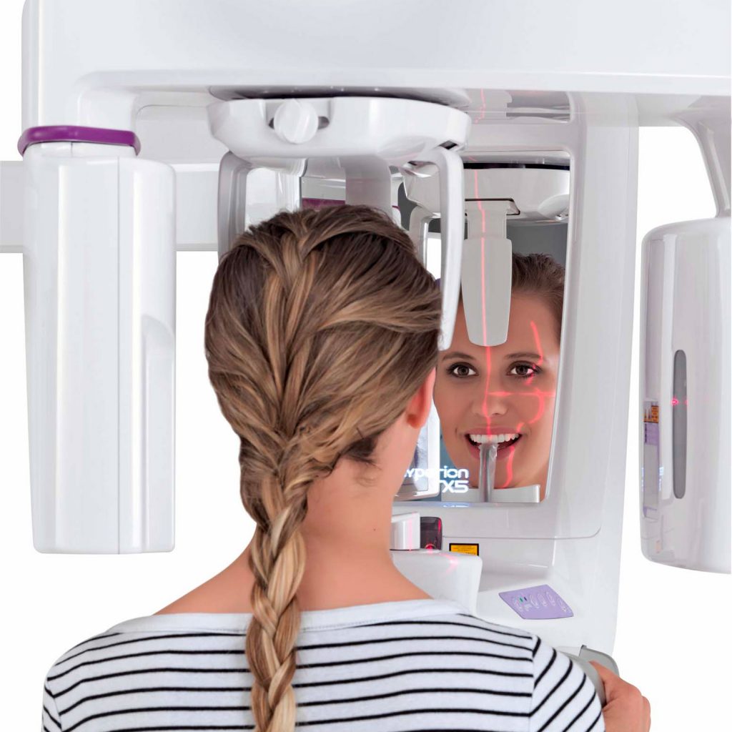
Achieving the full potential of 3D examinations has never been easier or more effective. Thanks to dedicated mechanisms, patient positioning solutions and exclusive automatisms that help ensure a positive outcome at every examination, dentists can make the most of 3D potential.
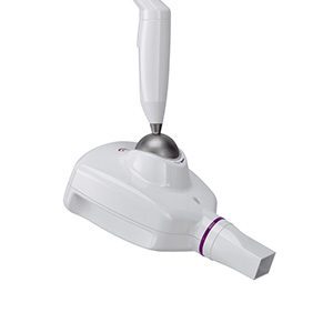
3d - PAN SENSOR
The high-sensitivity 3D sensor is also versatile as it can perform 2D panoramic imaging (managed by programs in the software package and controlled via the user-friendly virtual control panel).
ERGONOMIC HEAD SUPPORT
The dedicated head support for volumetric examinations has 5 contact points. An adjustable forehead support to improve patient positioning, two symmetrical side supports, which facilitate centring, a bite block and a chin rest, which guarantee the patient’s stability and, consequently, comfort and quality of the clinical examination.
AUTOMATIC CEPH COLLIMATION
In the event of cephalometric examinations the turret containing the 3D sensor is automatically rotated and lowered, aligning the opening integrated in the structure so as to create suitable collimation for the examination. Moreover, the sensor is positioned so as to make more space available for the patient and ensuring a more comfortable experience.
iRYS, simple and versatile diagnoses
The all-in-one software designed for simple and effective management of 2D and 3D images, with advanced tools and filters for diagnostics and planning.
CLINICAL CASES
2D
3D
CEPH
2D
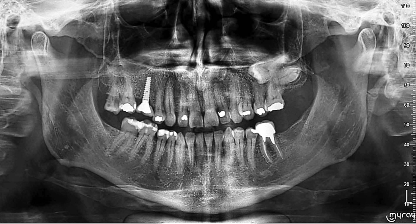
Adult Panoramic Imaging
Panoramic exposure programs calibrated on patient build to adapt X-ray doses accordingly.
Users can select the area of diagnostic interest for complete or partial analysis.
• QuickPAN or standard exposure
• Complete or partial analysis
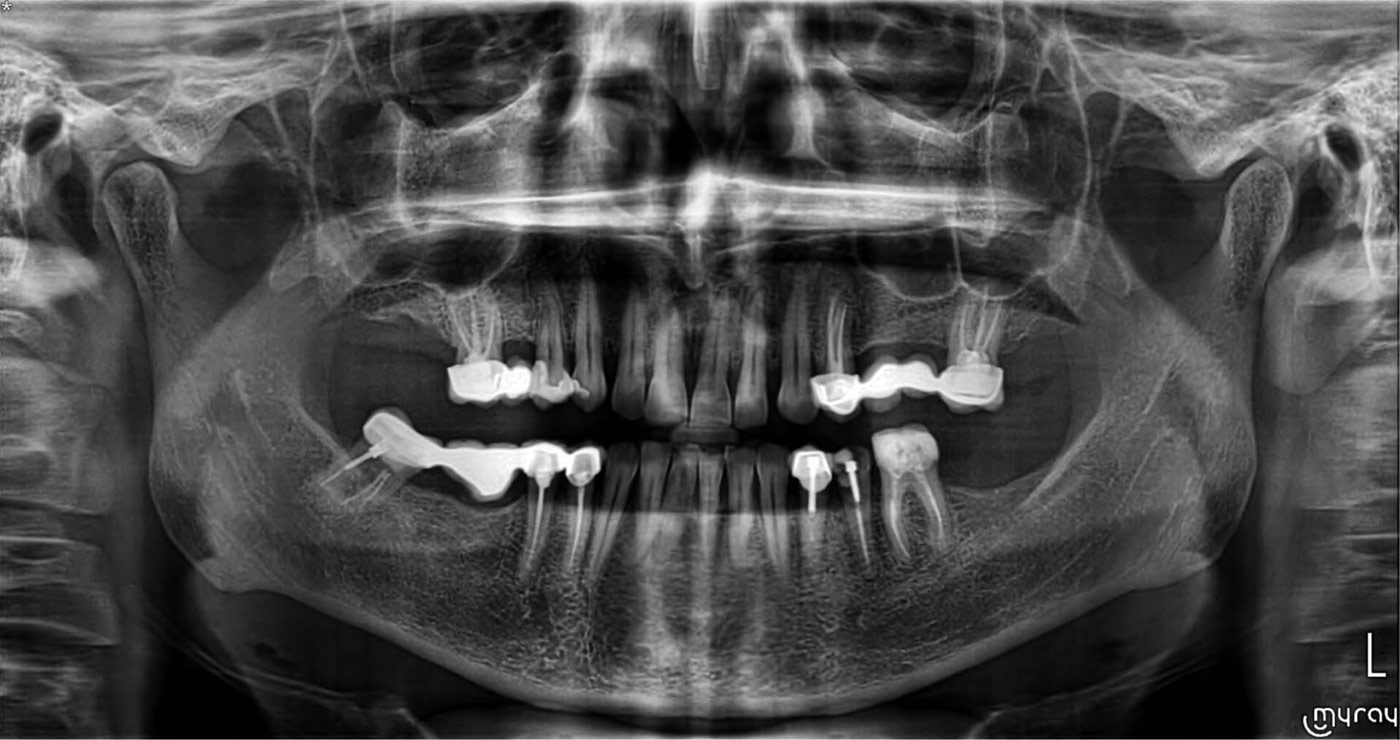
Orthogonal panoramic view
Minimises overlapping of adjacent tooth elements for improved periodontal examinations.
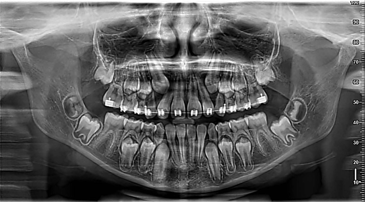
Child Panoramic Imaging
Limited exposure and optimised parameters for quick paediatric examinations. Users can select the area of diagnostic interest for complete or partial analysis.
• QuickPAN or standard exposure
• Complete or partial analysis
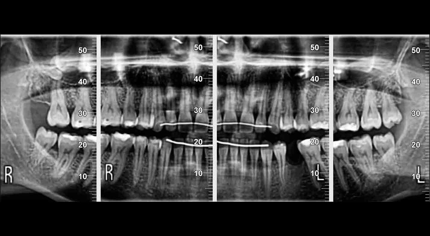
Dentition and Bitewing
Study of dentition with optimised interproximal projection for improved periodontal control. Collimation on the crowns for patients unable to tolerate intraoral bitewings, as they are more comfortable and less intrusive.
• Increased orthogonality
• Adapted collimation
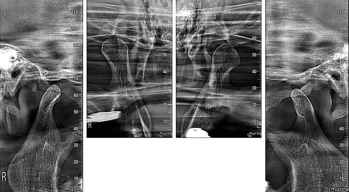
Temporomandibular joints
Simpler assessment of the temporomandibular situation thanks to latero-lateral or anteroposterior images, 4 X-rays in a single scan.
• Mouth open or closed
• Sagittal and Coronal
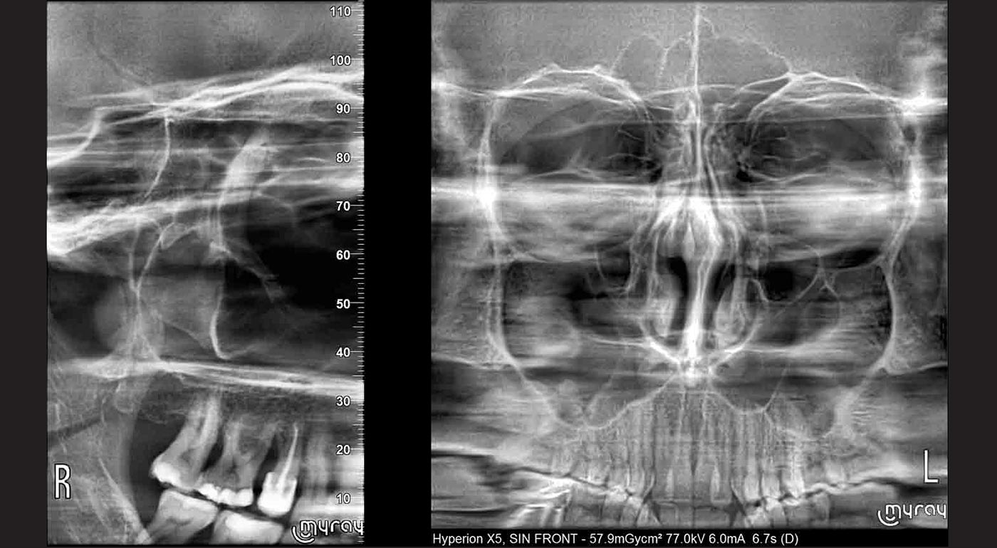
Maxillary sinuses
Characterised by a special image layer that provides X-rays in which the maxillary sinuses are clearly visible.
• Frontal
• Lateral
3D
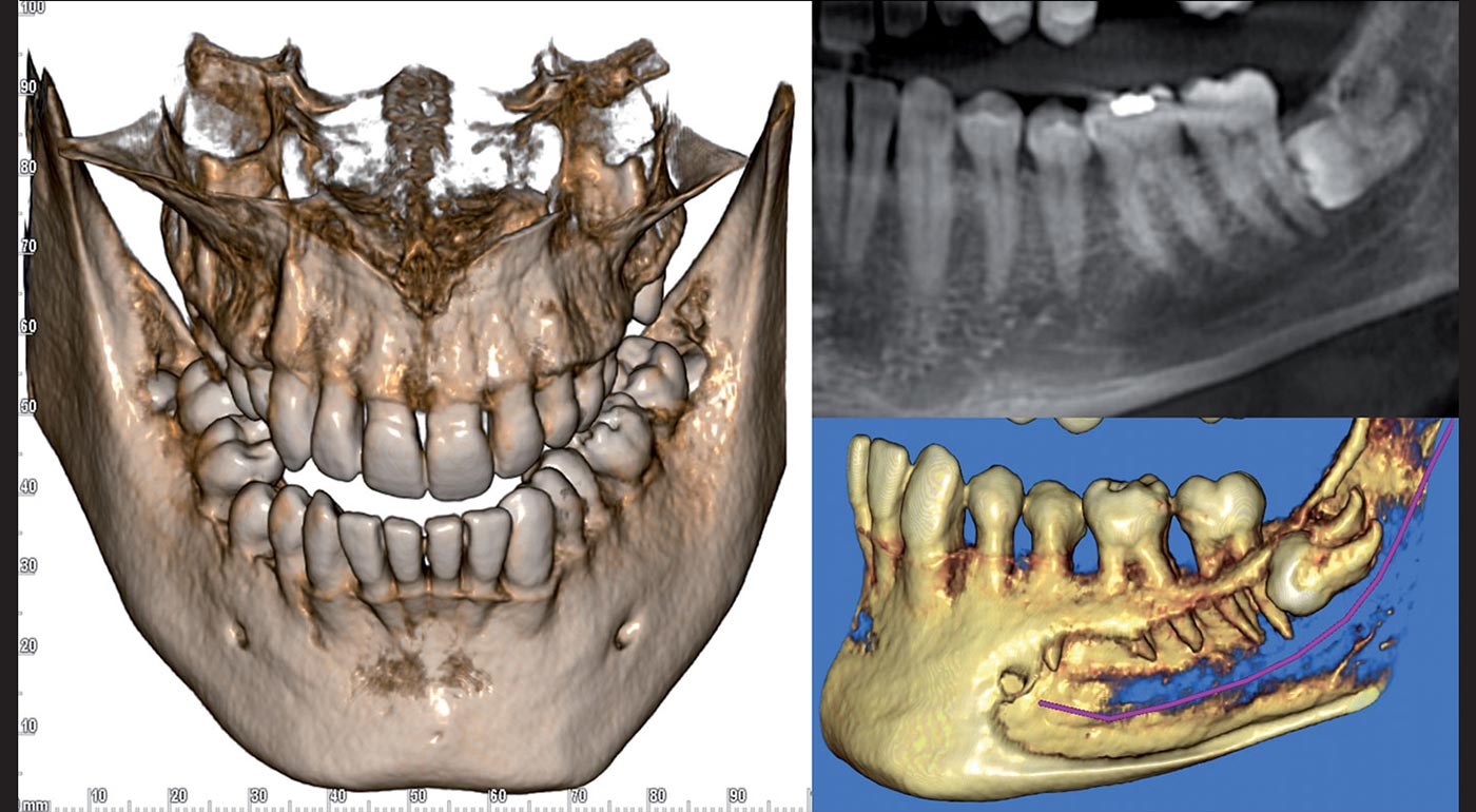
Complete (adult) dentition (adult)
Highly accurate investigation of both dental arches (including third molar roots) and surrounding anatomic features, useful for correct diagnosis and improved treatment planning. Unlike 2D, 3D allows identification of the true positioning identification.
• FOV 10 x 10cm with detailing up to 80 μm

Local (low dose) analysis
Diagnostics on the region of interest only, far more in-depth than 2D examinations, for HD endodontic assessments; study of relationship between impacted teeth; post-operation checks with fast scan and doses equivalent to those of a 2D examination.
• MultiFOV – HD and QuickScan
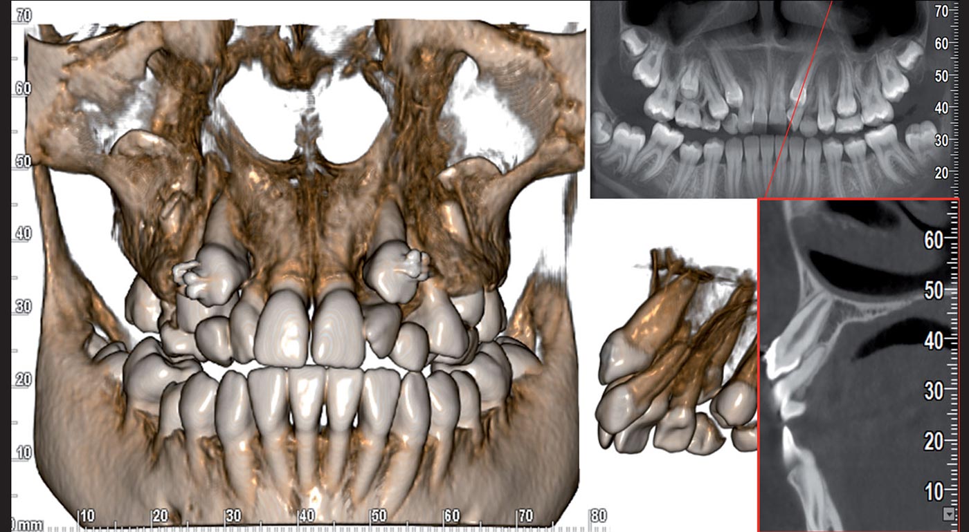
Complete (Child) dentition
Complete, low-dose volumetric examination of the dentition and maxillary sinuses of children. The reduced collimation avoids any exposure of sensitive organs while ensuring the investigation is complete and thorough investigation.
• Limited exposure – Low Dose
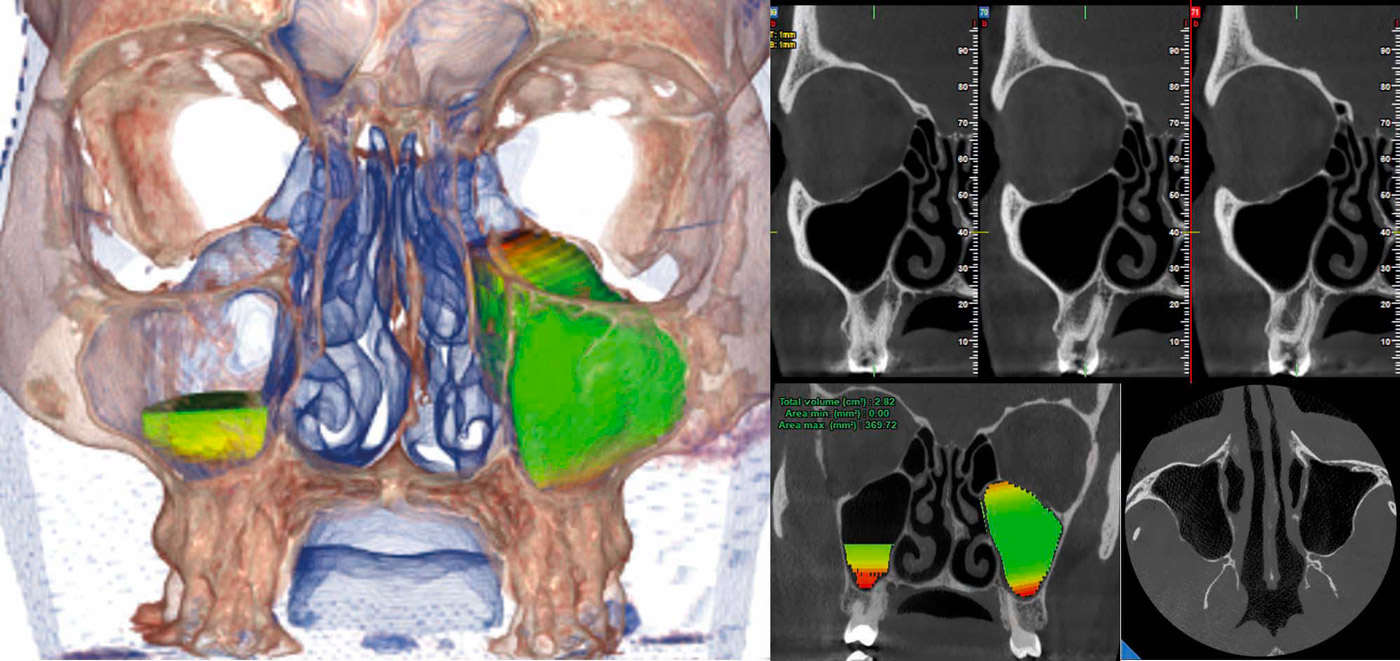
Maxillary Sinuses
The 10 x 10 cm FOV acquires in a single scan the maxillary sinus image useful for a volumetric assessment of structures and hollows. This allows any disease to be carefully diagnosed for optimised treatment planning, including sinus lifting, and volumetric analysis enabling to trace lines on a virtual patient model, evaluating morphological ratios on 3D renderings.
• Volumetric analysis – Low Dose
CEPH
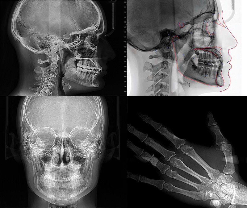
Teleradiography
Latero-Lateral: with highlighted soft tissue and bone details, critically important for cephalometric studies.
Anterior-Posterior: to detect asymmetries and malocclusions and be able to identify the right treatment.
Carpal bones: for residual growth potential assessment, possible with dedicated support.
Cefla Medical Equipment is considered Europe’s number one dental unit manufacturer.